The origin of image spliting in extreme blue and UV part of the spectrum is related to unfocused stellar image at the slit entrance because the presence of chromatic aberration. You can see image of telescope central obstruction in some circonstance.
The source of chromatism is double :
- the LISA internal aberration. Remember, the LISA optic is optimized for the wavelength range from 4000 A to 7100 A (for a f/5 equivalent telescope). The image deterioration increase more and more for wavelength shorter than 4000 A.
- the focal reduced added in front the LISA spectrograph is a major source of image quality degradation in the UV part. The majority of focal reducer are simple economic doublet, optimized for the visible part of the spectrum and poor UV quality. Try to remove the focal reducer and take an image for comparison.
Here a demo. First a focal reducer for an equivalent final beam at f/6.3 :
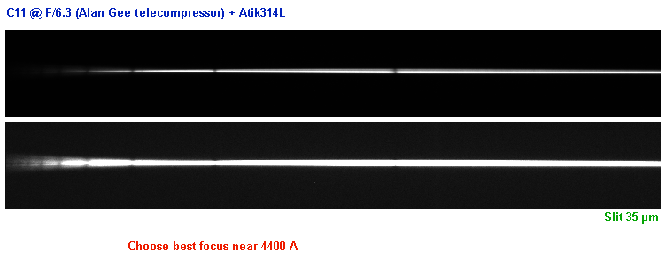
The spectrum quality is fine from 3950 A to 7100 A for scientific analysis.
Now the focal reducer is removed (f/10 input beam) :
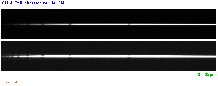
The quality is now fine down to 3800 angstroms (!). My LISA spectrograph is not perfectly adjusted by comparison to Emmanuel or Thierry models

Note : the best telescope for LISA is probably a Newton at F/5-F/7 (perfectly achromatic).
A key point is equalization of image sharpness only along the usable spectral range by focusing the star at the entrance slit.
Consider also the quality of slit image on the dectector surface (by adjusting the internal objective). Here a typical example of good image with my setup (internal neon lamp spectrum - 5 seconds exposure - 35 microns slit, the best choice for standard work):
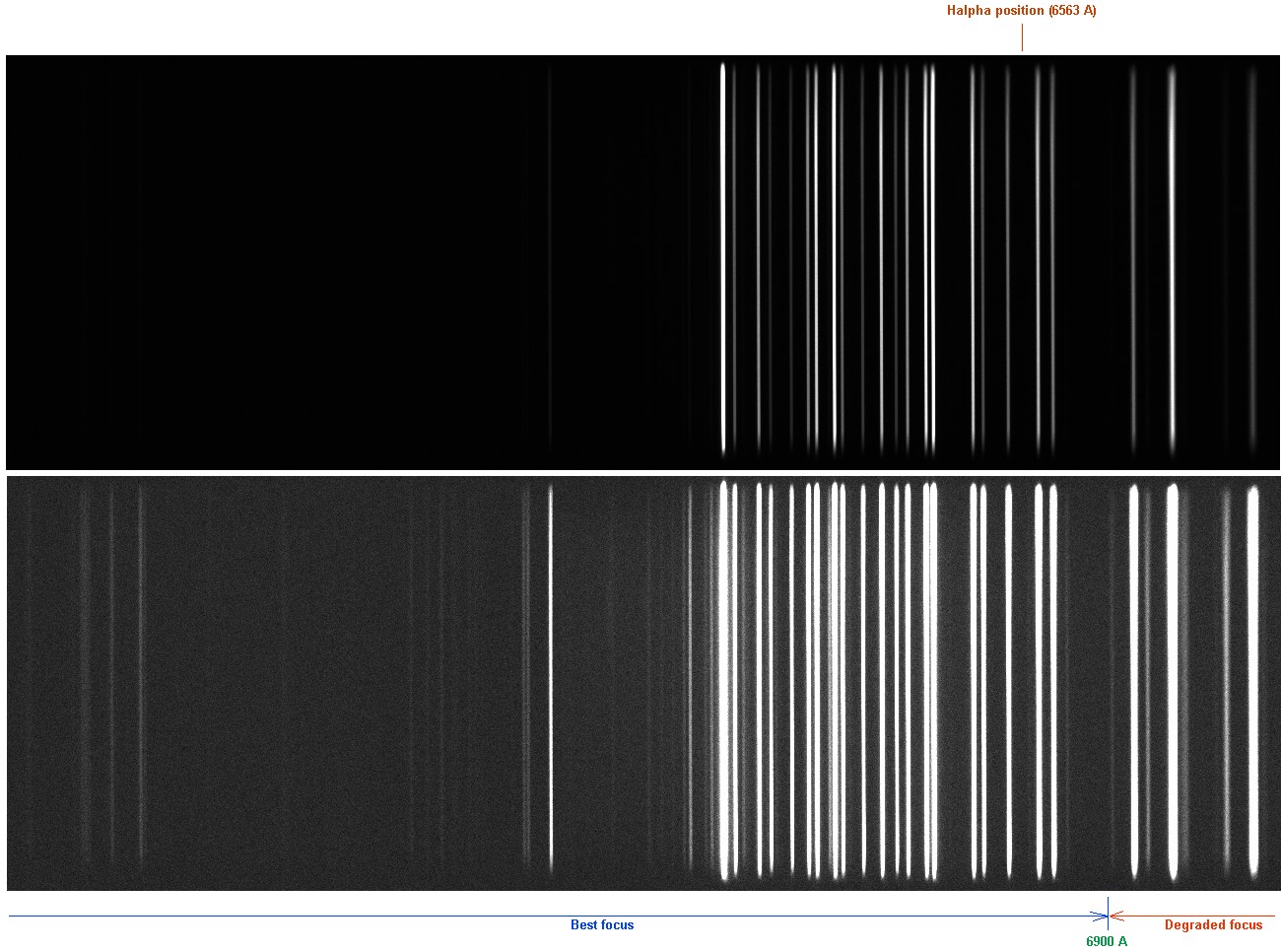
Do not attent to focus the IR part (of low astrophysic interest). Note the very sharp image at Halpha level. Try to reproduce this figure.
Christian B


